When your doctor orders a brain MRI, it’s not just a scan-it’s a window into what’s happening inside your brain. Unlike X-rays or CT scans, MRI doesn’t use radiation. Instead, it uses strong magnets and radio waves to create incredibly detailed pictures of brain tissue. This makes it the most powerful tool doctors have for spotting problems like strokes, tumors, multiple sclerosis, and even early signs of dementia. But what do those images actually show? And how do doctors tell the difference between normal aging and something serious?
What Brain MRI Actually Shows
Brain MRI doesn’t just take a picture-it captures how water behaves in different tissues. That’s why it’s so good at showing subtle changes. Healthy brain tissue has a clear balance of gray matter (where nerve cells live) and white matter (the wiring connecting them). On a T1-weighted image, gray matter looks darker than white matter, like a photo negative. Fluids like cerebrospinal fluid (CSF) appear black. That’s normal.
But when something goes wrong, water behavior changes. Swelling from injury or inflammation makes tissue hold more water, which shows up bright on T2-weighted and FLAIR images. That’s why doctors rely on these sequences to find lesions-tiny areas of damage that might be invisible on a CT scan. For example, in multiple sclerosis, plaques form where the protective coating around nerves breaks down. These show up as bright spots near the ventricles, the fluid-filled spaces in the brain’s center.
One common mistake is confusing normal age-related changes with disease. Nearly 90% of people over 70 have small bright spots on their MRI in the white matter. These are often just tiny blood vessel changes from years of high blood pressure or aging-not signs of MS or dementia. The key is location and pattern. Lesions in the deep white matter near the ventricles? Likely benign. Lesions in the cortex, brainstem, or spinal cord? That’s a red flag.
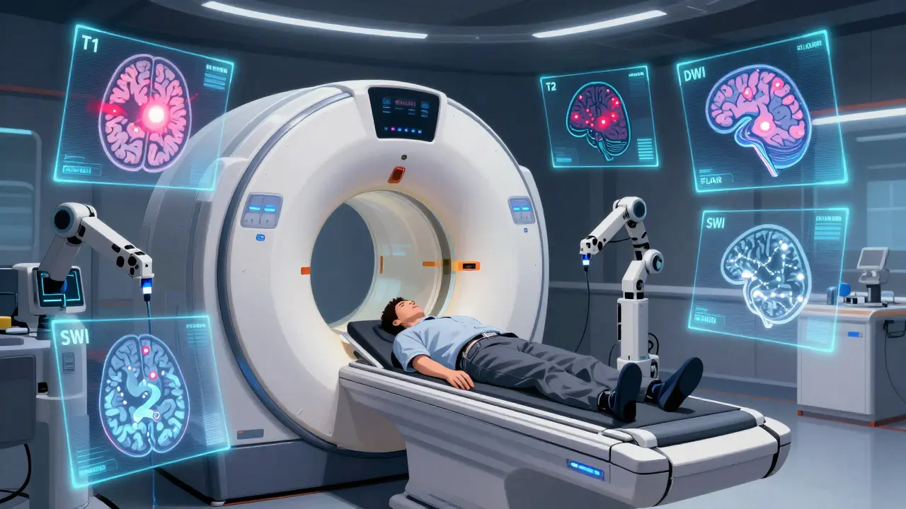
The Five Key MRI Sequences You Need to Know
Not all MRI images are the same. Each sequence highlights different things. Here’s what matters most:
- T1-weighted: Best for anatomy. Fat and some blood products look bright. CSF is dark. Used to see brain structure, tumors, and bleeding after a few days.
- T2-weighted: Shows water. Swelling, tumors, and inflammation glow bright. CSF is also bright, which can make it hard to tell if a bright spot is fluid or damage.
- FLAIR: The game-changer. It turns CSF black but keeps lesions bright. This is how doctors spot MS plaques, small strokes, and infections hiding near fluid-filled spaces.
- Diffusion-weighted imaging (DWI): The fastest way to catch a stroke. Within minutes of a stroke, water can’t move normally in brain cells. DWI shows this as a bright spot. If it’s bright on DWI but dark on ADC (the companion map), it’s an acute stroke-time-sensitive and treatable.
- SWI or gradient echo: Picks up tiny amounts of blood. Great for spotting microbleeds from high blood pressure, trauma, or amyloid angiopathy. These are often invisible on other sequences.
Doctors don’t look at one image-they compare all five. A bright spot on T2 that’s also bright on FLAIR? Probably a lesion. A bright spot on T2 but dark on FLAIR? Likely just CSF. DWI changes everything if you’re thinking stroke.
Common Findings and What They Mean
Here are the most frequent findings-and what they really mean in practice:
- White matter hyperintensities: Bright spots in the white matter. In people under 50, they’re uncommon. In older adults, they’re normal. But if they’re large, symmetrical, or near the brainstem, they could point to vascular disease, MS, or even rare genetic disorders.
- Small lacunar infarcts: Tiny (3-5mm) strokes deep in the brain, often in the basal ganglia or thalamus. These usually come from high blood pressure. They might not cause symptoms, but they increase stroke risk over time.
- Enlarged ventricles: Bigger fluid spaces can mean brain shrinkage (atrophy). This happens with aging, Alzheimer’s, or long-term alcohol use. But if the ventricles are enlarged and the brain looks normal, it could be normal pressure hydrocephalus-a treatable condition.
- Masses or tumors: Brain tumors show up as abnormal growths. Gliomas (from brain cells) often have blurry edges. Meningiomas (from the lining) are round and push the brain aside. Contrast dye helps tell them apart.
- Microbleeds: Tiny dots of old blood. Seen on SWI. Common in elderly people with high blood pressure. More than 5 microbleeds raises concern for amyloid angiopathy, a condition linked to Alzheimer’s.
- Acute stroke: Bright on DWI, dark on ADC. This is urgent. If caught within 4.5 hours, clot-busting drugs can help. Delayed treatment means permanent damage.
One thing to remember: finding something on an MRI doesn’t mean it’s causing your symptoms. A 70-year-old with memory issues might have dozens of white matter lesions-but the real cause could be vitamin B12 deficiency or thyroid trouble. That’s why doctors never read MRI alone. They combine it with your history, exam, and blood tests.
When MRI Isn’t the Right Tool
Despite its power, MRI isn’t perfect. For sudden head trauma, CT is faster and better at spotting skull fractures and fresh bleeding. If someone’s unstable, in pain, or can’t lie still for 40 minutes, CT wins.
Also, MRI can’t always tell if a lesion is new or old. A bright spot could be from a stroke last week-or ten years ago. That’s why contrast dye (gadolinium) is sometimes used. New, active inflammation lets dye leak in, lighting up the area. Old scars don’t. But even contrast has limits-it can’t always distinguish between tumor recurrence and radiation damage.
And here’s the hard truth: brain MRIs are often ordered for headaches with no other symptoms. Studies show only 1.3% of these scans find something serious. The American College of Radiology says MRI is usually not appropriate for uncomplicated migraines. Yet they’re still done-because patients ask, and doctors want to be sure. That’s why understanding what’s normal-and what’s not-is so important.
What Happens After the Scan?
After your scan, a radiologist spends 20-30 minutes reviewing every image. They compare both sides of the brain, check for symmetry, and look at each sequence. Then they write a report. That report doesn’t come with pictures. It says things like: “Multiple small white matter hyperintensities consistent with small vessel ischemic disease.” That sounds scary. But what it really means? “Your small blood vessels have been affected by aging or high blood pressure.”
Your doctor will explain what it means for you. If the MRI shows a tumor, you’ll be referred to a neurosurgeon. If it’s MS, you’ll see a neurologist for treatment. If it’s just age-related changes? You might get advice on controlling blood pressure, quitting smoking, or exercising more.
There’s no magic number of lesions that means “disease.” It’s the pattern, location, and your symptoms together that matter.

What’s New in Brain MRI?
Technology keeps improving. High-field 7.0T MRI machines are now in a few academic centers. They can show brain layers so clearly, scientists are mapping how signals travel between neurons. AI tools can now cut scan time in half-helping patients who can’t stay still, like those with Parkinson’s or dementia.
Quantitative MRI is coming fast. Instead of just saying “there’s a lesion,” future scans will measure how much water is in tissue, how fast blood flows, or how much myelin is left. These numbers could track disease progression better than ever-helping doctors know if a new drug is working before symptoms change.
But the biggest change isn’t the machine. It’s the understanding that not every bright spot needs treatment. The goal isn’t to find every tiny change. It’s to find the ones that matter-for your health, your life, and your future.
Can brain MRI detect Alzheimer’s disease?
MRI alone can’t diagnose Alzheimer’s, but it can show patterns that strongly suggest it. The most common sign is shrinkage (atrophy) in the hippocampus-the memory center of the brain. In Alzheimer’s, this shrinkage is usually symmetrical and gets worse over time. Doctors also look for thinning in the cortex and enlarged ventricles. For a definitive diagnosis, PET scans that detect amyloid plaques are used, but MRI is the first step. It rules out other causes like tumors or strokes that can mimic dementia.
Is a brain MRI safe if I have metal implants?
It depends on the implant. Pacemakers, cochlear implants, and some older aneurysm clips are absolute no-go’s because the magnet can move or heat them. But many modern implants are MRI-safe-especially joint replacements, dental fillings, and newer pacemakers labeled “MRI conditional.” Always tell the technologist about any metal in your body. They’ll check the device’s safety rating. If it’s unsafe, they’ll use CT instead. Never assume it’s okay-even a small tattoo with iron-based ink can heat up and burn you.
Why do I need to stay so still during the scan?
MRI works by detecting tiny changes in magnetic fields. Even a small movement-like swallowing or shifting your head-can blur the images. A 2mm shift can make a lesion look bigger than it is, or hide it entirely. That’s why you’re given pillows and straps. If you can’t stay still, the scan might need to be repeated, which adds time. For children or people with movement disorders, sedation is sometimes used. The goal is one clear set of images-not five tries.
Can brain MRI show anxiety or depression?
No. Anxiety and depression don’t show up as visible lesions or tumors on MRI. While research has found subtle differences in brain structure or activity in people with these conditions-like smaller hippocampi or altered connectivity-these aren’t reliable enough for diagnosis. Doctors don’t use MRI to diagnose mental health conditions. Instead, they rely on symptoms, interviews, and psychological tests. An MRI might be ordered if there are physical signs (like memory loss or seizures) that could point to a brain tumor or stroke mimicking depression.
How long does it take to get MRI results?
The scan itself takes 30 to 45 minutes. The radiologist usually finishes interpreting it within 24 to 48 hours. Your doctor will get the report and call you with the results. In urgent cases-like suspected stroke or sudden neurological decline-the radiologist will notify your doctor immediately, sometimes while you’re still in the scanner. Don’t expect results the same day unless it’s an emergency. Waiting isn’t a bad sign-it just means the team is being careful.
Are there risks from the MRI contrast dye?
The contrast dye used in brain MRI is gadolinium. It’s very safe for most people. Rarely, it can cause allergic reactions-like hives or nausea-but severe reactions are extremely uncommon. In people with severe kidney disease, gadolinium can lead to a rare condition called nephrogenic systemic fibrosis. That’s why kidney function is checked before giving contrast. Newer types of gadolinium are much safer. There’s no evidence it causes brain damage, even after repeated use. Still, doctors only use it when necessary-like when they suspect active inflammation, infection, or tumor growth.
What to Do Next
If your MRI shows something abnormal, don’t panic. Most findings are treatable-or even harmless. Ask your doctor: “What does this mean for me?” “Is this causing my symptoms?” “What’s the next step?”
If you’re healthy and your MRI came back normal, that’s good news. But don’t ignore lifestyle factors. High blood pressure, smoking, poor sleep, and inactivity all damage small blood vessels in the brain over time. Even if you don’t have symptoms now, protecting your brain starts today-with exercise, a healthy diet, and regular checkups.
Brain MRI isn’t magic. It’s a tool. Used right, it saves lives. Used wrong, it causes unnecessary worry. The best outcome isn’t a perfect scan-it’s understanding what you’re seeing, and what it means for your health.


8 Comments
doug b
Man, I had an MRI last year for migraines and got a report full of "white matter hyperintensities." My doctor just said, "You’re 42, you’ve had high blood pressure since college, and you’re not dead yet." Turned out it was just aging and bad coffee. No treatment needed. Just drink water, sleep more, and stop stressing about dots on a screen.
Irebami Soyinka
LOL 🤣 this whole post is like a medical textbook thrown into a TikTok algorithm. But hey, at least someone finally explained FLAIR without sounding like a robot. Nigeria has 3 MRI machines for 200 million people - we’re out here guessing strokes from eye twitching. Respect to the docs who actually know what they’re seeing. 🇳🇬💪
Katie Mccreary
So you’re telling me a 70-year-old with 20 bright spots on their MRI is fine, but if I have one at 35, I’m doomed? That’s not medicine, that’s ageism wrapped in radiology jargon. Also, why is gadolinium still being used if it’s "very safe"? My aunt developed brain fog after 3 scans. Coincidence? I think not.
Kevin Kennett
Biggest takeaway? MRI doesn’t diagnose you - your doctor does. I’ve seen people cry over "lesions" that turned out to be nothing. And I’ve seen people ignore symptoms because their scan was "clean." The machine doesn’t care about your life - you have to. Talk to your doc. Ask the hard questions. Don’t let pixels scare you.
Jess Bevis
Just flew from Nairobi to Chicago. Saw a guy with a pacemaker get turned away from the scanner. Told him, "In the US, we scan brains. In Kenya, we scan people." He laughed. Then cried. This post? It’s the bridge between those two worlds.
SRI GUNTORO
People are so careless with their brains. Smoking, sugar, no sleep, binge-watching Netflix until 3 AM - and then they panic when an MRI shows damage. You don’t get a free pass because you paid for a scan. Your lifestyle wrote that report. Own it.
Phil Davis
Interesting how we treat brain scans like horoscopes. "Oh, you have a bright spot? That means you’re stressed." Or "Your ventricles are enlarged - you’re basically already dead." Meanwhile, the real issue is you haven’t seen a therapist in 5 years and your cortisol is through the roof. MRI doesn’t measure burnout. But maybe it should.
Rose Palmer
Thank you for this meticulously structured and clinically accurate overview. The distinction between FLAIR and DWI sequences is particularly well-articulated, and the emphasis on clinical correlation over radiological findings aligns with current best practices in neuroimaging interpretation. I would only add that the increasing use of AI-assisted volumetric analysis may soon render subjective lesion counting obsolete - a development that promises greater objectivity and reproducibility in longitudinal tracking. Well done.